Dermatology
About dermatology
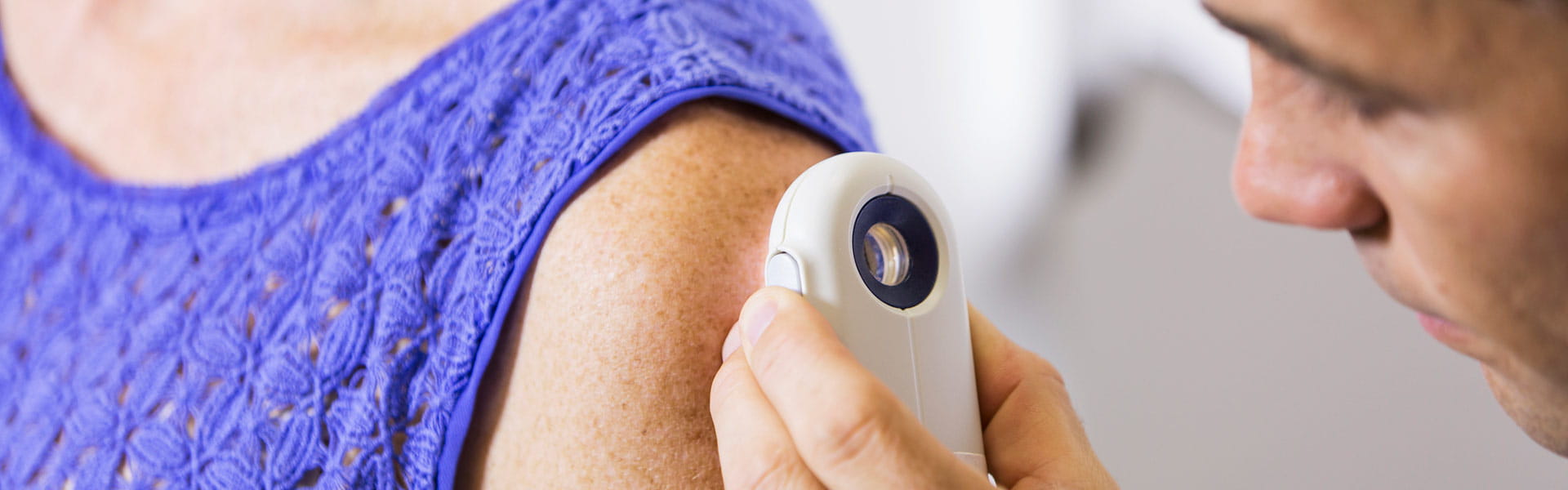
At Main Line Health dermatology, patients come to us with hair or skin problems and concerns about cancer and autoimmune disorders. We are also trained in cosmetic dermatology and provide treatments to improve the appearance of the skin, hair and nails.
Your skin is exposed to the environment, making it vulnerable to growths, rashes, cysts, burns and more. We see patients with a variety of conditions every day, including:
Our job is to address these issues so your skin can give you the best possible protection. We also work closely with Main Line Health experts of other specialties to bring you the precise, personalized solutions you need.
Acne
Aging Skin
Atopic Dermatitis or Eczema
Benign Skin Growth
Toenail Fungus
Contact Dermatitis
Hair Loss
Nail Disorders
Psoriasis
Rosacea
Scars
Skin Allergies
All Treatments
- Acne Treatments
- Dermabrasion
- Hair Loss Treatments
- Microdermabrasion
- Nail Treatments
Our Locations
Main Line Health Collegeville
Main Line Health King of Prussia
Bryn Mawr Hospital
825 Old Lancaster Road
Riddle Hospital
Lankenau Medical Center
Paoli Hospital
Main Line Health Newtown Square
Content you want, delivered to your inbox
Want to get the latest health and wellness articles delivered right to your inbox?
Subscribe to the Well Ahead Newsletter.